Reading Guide & Overview
Detecting Tissue With Qupath Information Center
Get comprehensive updates, key reports, and detailed insights compiled from verified editorial sources.
Get comprehensive updates, key reports, and detailed insights compiled from verified editorial sources.

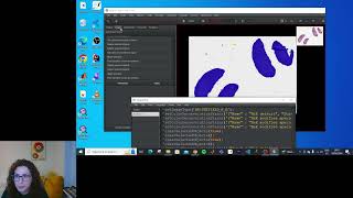
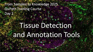
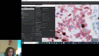
In this video, I show you how to separate stains in different types of images using This session of the FS2K workshop moves into the practical application of This video shows how to train a pixel classifier to segment epithelium and stroma in H&E images with The previous tutorial is at This time, we look at ... This tutorial walks you through the process of extracting high resolution images of individual cores from a In this tutorial is about pathology image annotation in
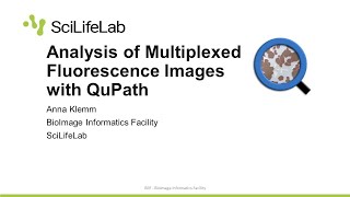
If you cannot read the text, make sure to turn the Quality up using the gear icon in the lower right corner of the video! This video shows how to train an object classifier for each fluorescent marker in a multiplexed image to identify positive cells for ...
Data is compiled from public records and verified media reports.
Last Updated: June 17, 2026
Stay updated on Detecting Tissue With Qupath's newest achievements.


For 2026, Detecting Tissue With Qupath remains one of the most talked-about profiles.
Below is a handpicked selection of video coverage regarding Detecting Tissue With Qupath.

Explore the primary sources for Detecting Tissue With Qupath.
Disclaimer:


